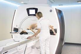
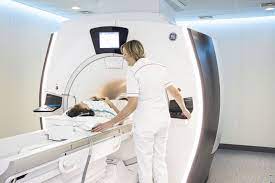
MRI imaging

The history of brain MRI in MS began on November 14, 1981, when an article entitled “Nuclear magnetic resonance imaging of the brain in Multiple Sclerosis” was published in the journal “The Lancet” by Y. R. Young and collaborators. In it, the authors showed that in 10 patients with MS, the X-ray scanner (CT-scan) revealed only 19 lesions in total, while the MRI revealed 112 additional lesions!
It was therefore the first time that it was possible, during the lifetime of a patient, to realize the extent of the disease and the number of brain lesions of which he was a carrier (“his lesional burden”). We quickly realized that most of these lesions were asymptomatic, in so-called “mute” areas of the brain.
The MRI then gave us the opportunity to distinguish between old lesions and active inflammatory lesions.
Significant improvements have been made to the quality of the images obtained, to their resolution, thanks to the increase in the power of the magnets used (0.5 then 1.5 then 3 Tesla), and to the adaptation of this technique to the spinal cord. It then gave us the possibility of distinguishing old lesions from active inflammatory lesions (new lesions or reactivation of old lesions) where there is a break in the hematoencephalic barrier. Active lesions in fact capture a paramagnetic contrast product called gadolinium. An active lesion can evolve (but not systematically) into a central necrosis called “black holes”, due to a destruction not only of the myelin sheaths but of the nerve fibers.

MRI was used as early as 2001 in the criteria for diagnosing the disease based on the number and location of lesions, either periventricular, cortical or juxtacortical, or in the cerebellum, brain stem or spinal cord. Finally, it demonstrated what neuropathology had already taught us, namely the presence of a central vein around which most lesions form. On the other hand, the subpial demyelination areas, just below the meninges, are still difficult to detect by conventional MRI.
It was possible to define prognostic criteria for the disease on the basis of the images observed. Thus, lesions located in the brain stem, cerebellum, or lateral cords of the spinal cord have a poorer prognosis than injuries that are only periventricular and hemispheric. Some lesions (but not all) were detected within the cerebral cortex in addition to those located in the white matter containing the myelinated fibers. It is now possible to measure brain atrophy induced by the disease beyond the normal decrease in brain volume of up to 0.4% per year. There are also more selective and localized atrophies, for example, in the corpus callosum, which contains nerve fibers connecting the 2 cerebral hemispheres, the thalamus and the cervical medulla. More recently, MRI revealed foci of focal meningitis corresponding to lymphocyte nodules in the meninges.
Slowing this brain atrophy down to normal values observed in any person was the aim.
It is also thanks to MRI that we were able to show the partial but significant effectiveness of the first interferon used in the disease, Betaferon, by proving its effectiveness not only by reducing clinical relapses but also by reducing the number of new lesions. This first study, published in 1993, did not yet use contrast medium (gadolinium), which was the rule in later studies. Global brain atrophy has also become another measure of drug effectiveness, used for the first time in the fingolimod (Gilenya) study. Slowing this brain atrophy down to normal values observed in all people has been the aim sought and achieved in several recent studies of new treatments.
Until now, conventional MRI has not been very quantitative in terms of the overall volume of lesions and in terms of the presence of active chronic lesions evolving at low noise without breaking the hematoencephalic barrier. However, these latter lesions are very important in the progressive forms of the disease.
More recently, it was possible to show that they were partially or totally surrounded by a thin line of iron-containing inflammatory cells. However, as Dr. Solène Dauby discusses, this technology can still provide us with new elements in the knowledge of the disease, for example by using a magnet more powerful than 7 Tesla. It will make it possible to analyze and quantify nerve cell losses, the rarefaction of synaptic connections, the decrease in the density of nerve fibers, and potentially, the remyelination of some of them, spontaneously or thanks to new treatments under experimentation.
Prof. Dr. Christian Sindic