Scientific
newsletters

MRI, a crucial tool for the diagnosis, prognosis and monitoring of patients
Multiple sclerosis is often described as an invisible disease or the disease of a thousand faces. MRI plays an important role in the diagnosis of this currently incurable disease. It can be used to confirm raised suspicions in addition to demonstrating the exact location and number of lesions. Dr. Solène Dauby explains ...
Currently MRI plays a major role in the assessment and monitoring of patients with MS. It is a crucial tool for the diagnosis, prognosis and monitoring of these patients The changes that this disease induces in brain tissue are very complex and include inflammatory cell infiltration, the destruction of certain components of nerve cells (axons, synapses, etc.) and their sheaths (myelin), neuronal death, hypertrophy and the activation of other brain cells (astrocytes, microglia, etc.). Although we are unable to see these cells in detail, MRI can detect some of the secondary lesions associated with these phenomena. (1)
What determines the severity of the disease?
This is a question that I often get from patients. It is quite easy and even tempting to imagine that a large number of lesions and substantial cerebral volume loss imply that the disease is more severe. It is not as easy as that, however. Observations have shown that the correlation between the number of lesions observed on conventional MRI (to which we have access in the clinic) and the disability in some patients is imperfect. In my opinion, there are two important reasons for this:
IIt is clear that the location of the lesions is a very important factor in determining the consequences that the lesion may have in terms of disability
Firstly, the brain is organised into different areas and the functionality of each of these areas is very different. Obviously, the location of the lesions is a very important criterion for the lesion’s potential impact in terms of disability. A small lesion, in an important area of the brain (for example, used to move a limb), will have a much greater clinical impact than a larger lesion that is located in a more ‘silent’ area.
Secondly, conventional MRI only reveals part of the pathological processes of MS, and that is why we conceived this project. When we compare the MS lesions with MRI and then, under the microscope after autopsy, we observe several tissue changes that are linked to the disease but that are not visible with conventional MRI. Conceivably, the clinical condition and disability of a patient could thus worsen even though the MRI remains unchanged! This is probably due to these subtle changes, which conventional MRI cannot detect, but which are occurring in the brains of our patients. This is consistent with what we observe in progressive forms of this disease.
Are there other imaging techniques that can be used to identify MS (now and in the future)?
There are other imaging techniques that are used to study MS, but these are mostly being tested for research purposes. We also intend to use PET scans (2), more specifically to study the synapses as part of our study. But in clinical practice, MRI is used for the most part for clinical monitoring and to evaluate the effectiveness of a treatment. Besides imaging techniques, we can also use clinical tests and objective measurements of specific parameters to visualise and assess this disease.
Does MRI also play a role in determining whether a patient should change treatments?
Yes, of course. In recent years, the management of MS has undergone significant changes, thanks to the advent of immunomodulatory therapies. Several studies have demonstrated that the functional prognosis (i.e. the patient’s autonomy, his ability to walk, move, speak, etc.) is better when the disease is managed as early and as effectively as possible.
MRI monitoring is crucial to managing patients as effectively as possible. We should therefore be able to adapt the treatment in step with the disease’s potential aggressiveness. It is not uncommon to start a treatment and then switch to a more effective therapy because the disease progresses and new relapses eventually occur. Regular MRI follow-up can detect when treatment needs to be stepped up at the earliest possible stage.
Your research, which is funded by the Charcot Foundation, aims to clarify the correlation between what the MRI shows and the natural progression of the disease... Can you explain how you intend to do this?
As mentioned earlier, one of the current challenges in the treatment of MS is this imperfect correlation between lesions on conventional
MRI and the patient’s condition or progression. We know that conventional MRI does not detect everything and that we therefore miss certain phenomena, certain lesions
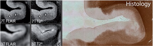
They explain this discrepancy. Our research project aims to use two very advanced imaging techniques, namely 7T MRI (3) and PET scan. These two technologies will hopefully give us access to specific lesions and abnormalities that are not visible with conventional MRI.We hope that by increasing our ability to detect brain abnormalities in patients, we can better correlate the images and clinical observations.
Source: Kilsdonk et al Brain 2016. This image shows that 7T MRI can approach the anapathic appearance of cortical lesions.
A new line of research focuses on remyelination. Can you explain how MRI or other imaging technology might contribute to this research?
A number of specific parameters that are studied using quantitative MRI (specific protocols used for MRI in research) have a correlation with the amount of myelin. Recently, researchers demonstrated that when you measure a quantitative parameter in MS patients called magnetisation transfer or MT, you can observe a correlation between the evolution and clinical data. In patients with a favourable course, the MT parameter in the lesions tends to increase.
Dr. Solène Dauby
(1) MRI is a medical examination performed using strong electromagnetic fields. It produces 2D and 3D images of the body.(2) PET scan is an imaging technique that studies the metabolic activity of tissues using a radioactive tracer like or analog to glucose. (3) 7 Tesla MRI is a pioneering technique that provides enhanced images which can be used to map areas in the brain to the level of infra-millimetre resolution. Recently, 7T MRI demonstrated that MS, which until then was considered to attack the white matter of the brain only, is in fact also accompanied by lesions in the grey matter, which cannot be detected on conventional MRI.

Newsletter 50
Stay informed
Receive all the information related to research and news from the Belgian Charcot Foundation directly in your inbox.